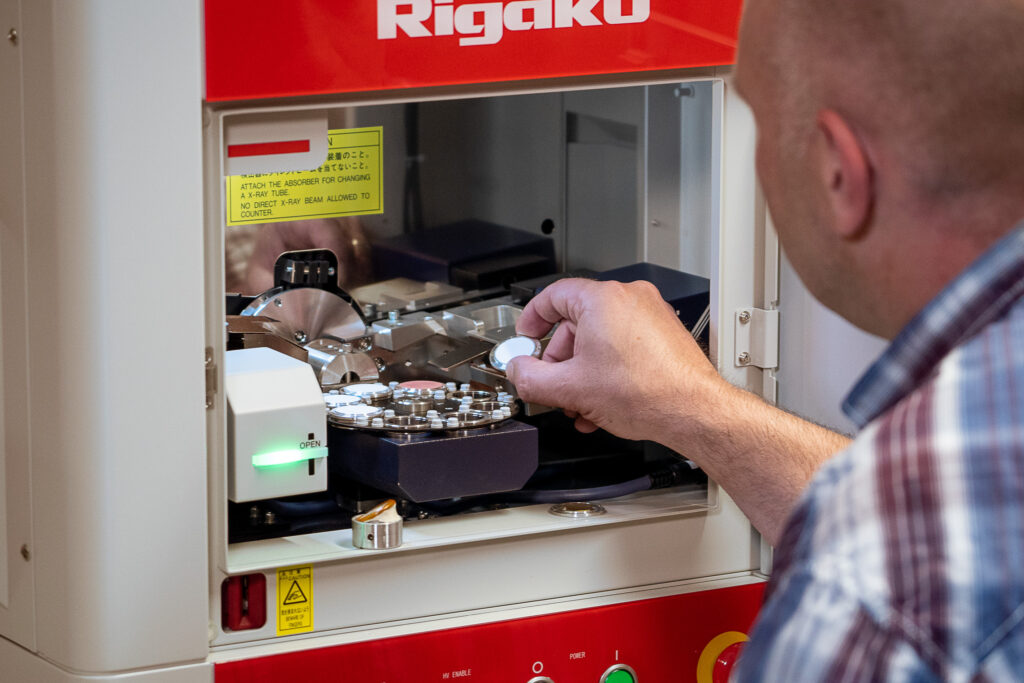
X-ray Structure Analysis

The Principle Briefly Explained
X-ray Structure Analysis
X-ray structure analysis allows the identification of crystalline materials via X-ray diffraction (XRD) on the crystal lattice. The position and intensity of the maxima in the diffraction pattern depend on the arrangement of atoms in the crystal lattice and are thus specific to a material.
X-ray structure analysis is usually performed on fine powders, which is why it is also called powder diffractometry and serves to:
- Identification of crystalline solids and their quantification
- Determination of crystal modifications of a compound (phase analysis)
- Measurement of lattice parameters, crystallite sizes, and degree of crystallinity
- Characterization of hydroxylapatite regarding crystallinity, phase purity, and Ca/P ratio according to ISO 13779-3
Application Examples
Phase Purity with X-ray Diffraction
X-ray Diffraction – Purity Analysis of Bone Substitutes
Problem Statement
The mineral hydroxylapatite – Ca5(OH)(PO4)3 – is a main component of human bone substance and has proven itself as an implant material for surgical applications.
Calcium compounds exist with a similar chemical composition but with a different crystal structure and consequently altered or undesirable properties regarding biocompatibility or resorption rate.
Solution
X-ray diffraction allows the identification and quantitative detection of foreign phases.
For the example shown in Figure 1, the hydroxylapatite contains traces of calcium oxide (marked by reflections with arrows).
The method is described in standard ISO 13779-3.
Another important property that can be determined by X-ray diffraction is the degree of crystallinity of the sample.
Amorphous components demonstrably have higher solubility and can be resorbed more quickly in the body.
An evaluation of the peak width in the diffraction pattern allows conclusions about crystallite size.
The size and shape of hydroxylapatite crystals (HA) are additionally investigated using electron microscopy (see Fig. 2).
Industries & Applications
- Medical Technology
Objectives
Product Development, Quality Assurance, Failure Analysis
Materials
Crystalline Solids, Bone Cements
Analysis Methods
- X-ray Diffractometry (XRD)
- Wide-Angle X-ray Scattering (WAXS)
Complementary Methods
- X-ray Fluorescence (XRF)
- Electron Microscopy
Advantages
X-ray diffraction allows statements regarding phase purity, crystallinity, and crystallite size. These are parameters that must be checked for quality assurance according to standards. In addition to the application example from medical technology, this technique can also distinguish various modifications of the white pigment titanium dioxide or various calcium sulfates (gypsum, bassanite, anhydrite, etc.). For the analysis of hydroxylapatite, Analytik Service Obernburg GmbH also offers the possibility to analyze the Ca:P ratio and heavy metal freedom by X-ray fluorescence (XRF). The ICP-OES method allows the analysis of heavy metal impurities even in the lowest concentrations.

Figure 1: Diffraction pattern of a powder sample plotted as intensity versus diffraction angle (blue line, top). For comparison, below are reflection positions and intensities from a database for hydroxylapatite (green) and calcium oxide (red).

Figure 2: Visualization of the needle structure and size of hydroxylapatite crystals using transmission electron microscopy.
